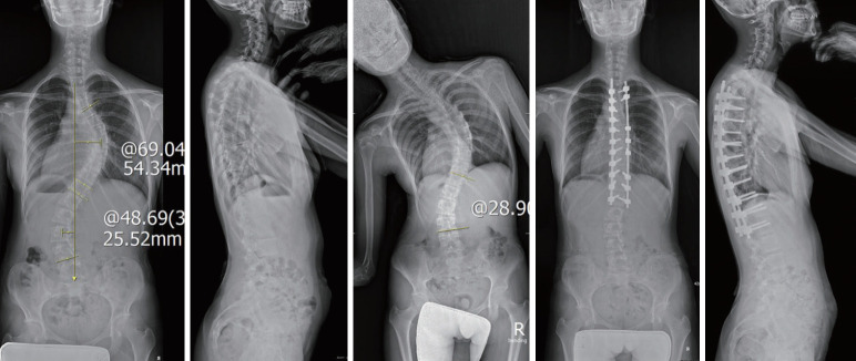
Fig. 2.
A representative case of selective thoracic fusion for Lenke 3CN curves. Preoperative standing posteroanterior radiography showed a right-side major thoracic curve of 69.0° and a left-side structural lumbar curve of 48.6°, which was reduced by 28.9° on the side bending film. The thoracic apical vertebral translation (AVT) was 54.3 mm and the lumbar AVT was 25.5 mm. Considering that the upper end vertebra was T5 with level shoulders, T3 was chosen as the upper instrumented vertebra. The lower instrumented vertebra was located at L1, which was the last substantially touched vertebra by central sacral line. Therefore, the patient underwent selective thoracic fusion from T3 to L1.

