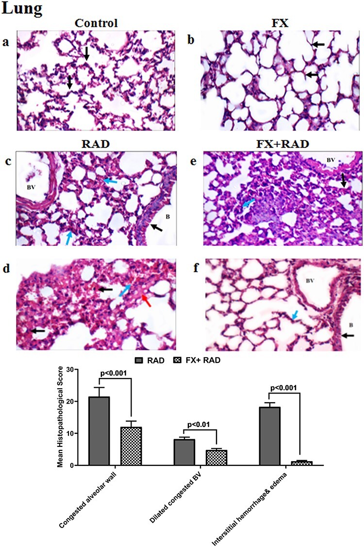
Fig. 9.

Histopathological examination of the lung in different animal groups. All tissues sections are stained with haematoxylin and eosin, magnification ×400 (H&E ×400). Control group, normal mice; RAD group, mice exposed to γ-radiation; FX group, mice treated with fucoxanthin; and FX + RAD group, mice treated with FX and exposed to γ-radiation.
