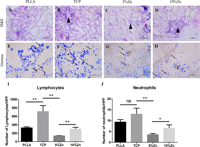
Fig. 3.
The moderate local inflammatory reaction elicited by scaffolds at the beginning of grafting in vivo. H&E (scale bar = 100 μm) (A–D) and Giemsa (scale bar = 50 μm) (E–H) staining of the scaffolds implanted subcutaneously in rats for 3 days, respectively. Statistical semiquantification of lymphocyte (I) and neutrophil (N) invasion of histological sections from the implanted scaffolds. PLLA, PLLA scaffold; TCP, TCP/PLLA scaffold; 5%Zn, 5%Zn-loaded TCP/PLLA scaffold; 10%Zn, 10%Zn-loaded TCP/PLLA scaffold; ns, no significance; *p < 0.05; **p < 0.01

