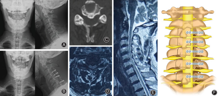
Fig. 1.

(A) Preoperative DX showed that the physiological curvature of the cervical spine became straight. (B) Postoperative DX showed that the internal fixation position was good. (C) CT showed C4‐5 level spinal stenosis. (D) MRI cross‐section showed C4‐5 level spinal stenosis. (E) MRI sagittal plane showed cervical spinal stenosis. (F) Schematic illustration.
