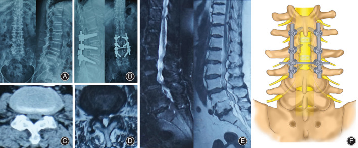
Fig. 2.

(A) Preoperative DX showed that the physiological curvature of the lumbar spine was straightened; (B) Postoperative DX showed good internal fixation position; (C) CT shows L3‐4 spinal stenosis; (D) MRI cross section shows L3‐4 spinal stenosis; (E) MRI sagittal plane shows lumbar spinal stenosis. (F) Schematic illustration.
