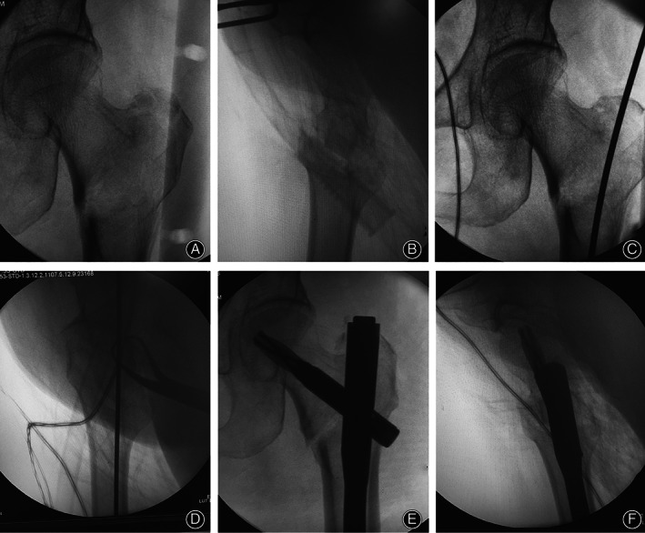
Fig 2.

Intraoperative fluoroscopy images. Anteroposterior (A) and lateral (B) radiographic images of closed reduction facilitated by double reverse traction repositor. Quality of reduction is accepted as “excellent.” Anteroposterior (C) and lateral (D) radiographic images of the guide wire insertion. Anteroposterior (E) and lateral (F) radiographic images of the PFNA‐II nailing demonstrated that the anatomic reduction was maintained by the double reverse traction repositor and that the fixation quality was optimal.
