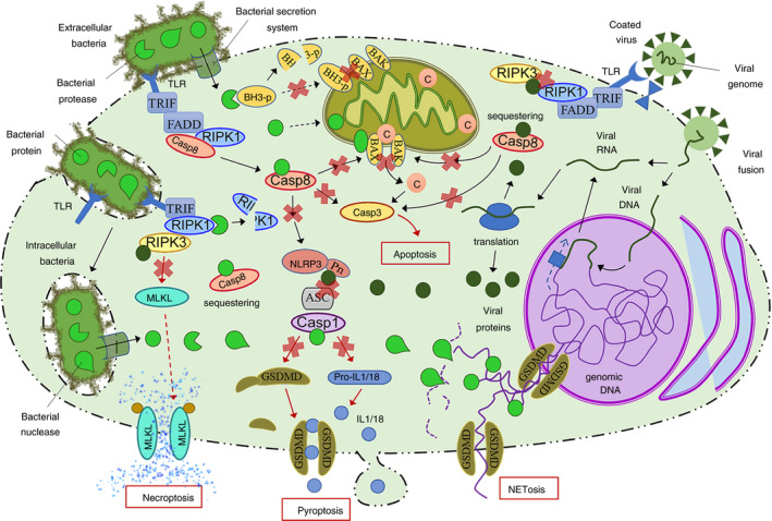
FIGURE 2.

Overview of the main bacterial and viral mechanisms to suppress PCD such as apoptosis and major forms of programmed necrosis. Both extracellular and intracellular bacteria have developed pore‐forming secretion systems to deliver specific proteins inside the cell to inhibit the main targets of multiple processes including cell death. Bacterial suppression of PCD is based on proteases and inhibitory proteins that decrease the integrity/activity of host cell factors. Some bacteria can also produce nucleases and DNA binding proteins that impair NET formation. On the other hand, viruses (including DNA and RNA viruses) deliver their genetic material into the cell where it is processed by the host cell machinery to undergo transcription and translation or direct translation, leading to the expression of viral proteins. These virulence factors suppress PCD mainly interacting with target proteins to sequester them or occluding binding domains to impair protein–protein interaction. Black arrows represent sequential steps in a process/pathway, and red arrows represent the final steps in a given pathway. Red crosses represent inhibition or blockade of a protein/pathway. ASC, apoptosis‐associated speck‐like protein; BH3‐p, BH3 only domain‐containing proteins; C, cytochrome c; Casp, caspase; FADD, Fas‐associated protein with death domain; GSDMD, Gasdermin D; IL, interleukin; MLKL, mixed lineage kinase domain‐like protein; NET, neutrophil extracellular trap; NLRP3, Nod‐like receptor pyrin‐containing protein 3; Pn, pyrin domain; RIPK, receptor‐interacting protein kinases; TLRs, Toll‐like receptors; TRIF, TIR domain‐containing adapter‐inducing interferon‐β
