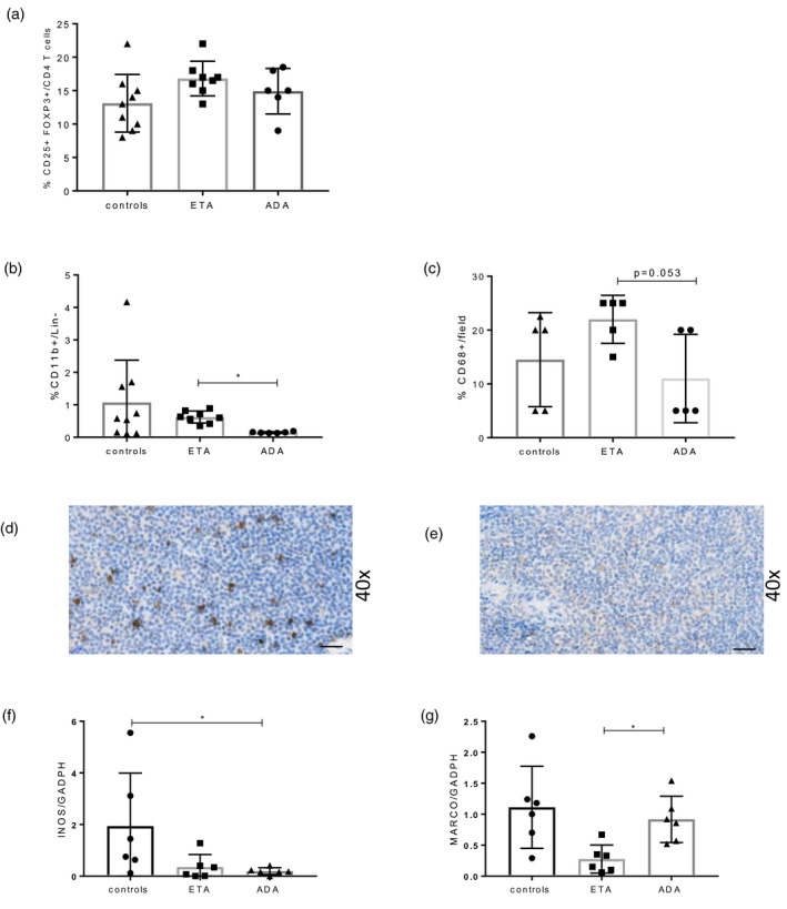
FIGURE 6.

Assessment of cellular infiltrate involved in lymphoma immunosurveillance in tumor necrosis factor (TNF) family‐transgenic (BAFF‐Tg) mice treated with TNF inhibitor (TNFi). Flow cytometry analysis was performed on spleen of BAFF‐Tg mice treated by control (n = 9), eternacept (ETA) (n = 8) or adalimumab (ADA) (n = 6). (a) Splenic regulatory T cells (Treg) and (b) macrophages were assessed. (c) Macrophage infiltrate was also assessed by CD68 staining in immunohistochemistry (IHC) on spleen sections (n = 5 for each group). Panels (d) and (e) are representative of CD68 staining in IHC, with ×40 magnification in mice treated with (d) ETA and (e) ADA. Inducible nitric oxide synthase (INOS) (f) and MARCO (g) in ADA (n = 6), ETA (n = 6) or control immunoglobulin (Ig) mice (n = 6) quantified by quantitative polymerase chain reaction (qPCR). Results are shown as mean and standard deviation (s.d.). Kruskal–Wallis test followed by Dunn’s multiple comparison test. *p < 0.05, **p < 0.01
