
Fig. 3.
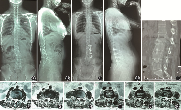
A typical case of a 58‐year‐old female who underwent a combined OLIF and UPSF for correction of DLS. (A) Anteroposterior (AP) view of preoperative standing full‐length spinal radiograph showed scoliosis. The Cobb angle was 30.8 degrees. (B) Lateral view of preoperative standing full‐length spinal radiograph showed sagittal alignment of the patient. (C) AP view of postoperative standing full‐length spinal radiograph showed correction of scoliosis. The Cobb angle was 0.3 degrees. (D) Lateral view of postoperative standing full‐length spinal radiograph showed realignment of sagittal plane. (E) 12‐month postoperative CT showing good alignment and fusion status. (F, G, H) The preoperative axial T2WI MRI scan showed narrowing of the spinal canal of L2–L3, L3–L4, and L4–L5. (I, J, K) The postoperative axial T2WI MRI scan showed indirect decompression of the spinal canal of L2–L3, L3–L4, and L4–L5.