
Fig. 5.
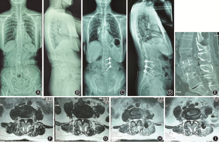
(A–D) Pre‐ and postoperative standing full‐length spinal radiograph of patient who underwent a combined OLIF and UPSF for correction of DLS. OLIF procedure was performed at L3–L5 where degenerative change of intervertebral space happened. However, the coronal and sagittal spine alignment has been improved. (E) Interbody fusion had occurred. (F, G) Preoperative axial T2WI MRI showed narrowing of the spinal canal of L3–L4 and L4–L5. (H, I) Postoperative axial T2WI MRI showed indirect decompression of the spinal canal of L3–L4 and L4–L5.