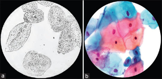
Figure 2.

Evolution of medical documentation via the photomicrograph. One of the first applications of photography was that of the photomicrograph, as illustrated in Alfred Donne's Atlas du Cours de Microscopie, and entitled “Epidermal cells of normal vaginal mucosa” (translated) in 1845 (a, public domain).[6] Today, photomicrographs are still utilized to document and convey vital concepts in anatomic pathology (b, ThinPrep with Papanicolaou stain, ×600 original magnification)
