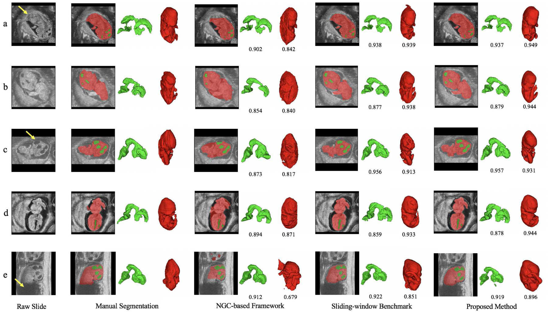
Fig. 7.

Comparison of qualitative segmentation results among different methods for five HFU images. Green indicates BV, red indicates body and the numbers below the predicted segmentation are corresponding DSC. Yellow arrow in a) indicates ambiguous boundary due to the deep touching of the body and uterine wall. Image b) has severe motion artifacts. Yellow arrow in c) indicates missing head boundary. Image d) has different contrast with image b) and c). Yellow arrow in e) indicates severe missing signal of body, which leads to unsatisfactory automatic body segmentation results across different methods.
