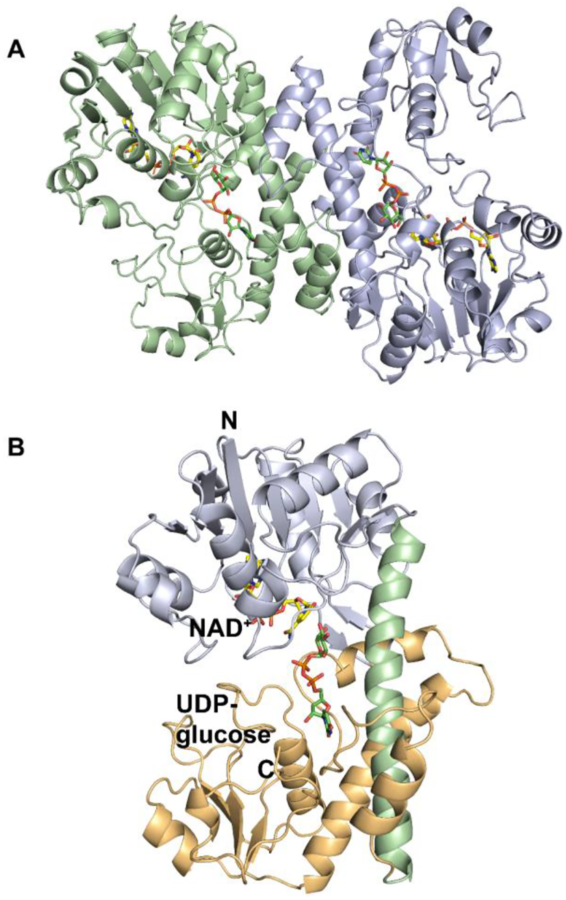
Figure 5.

(A) Ribbon diagram of Cj1441 homodimer. Chain A is shown in pale green, and Chain B is shown in pale blue. UDP-glucose is depicted in green and NAD+ is displayed in yellow. (B) Ribbon diagram of Cj1441 monomer where the N-terminal domain is pale blue and the C-terminal domain is shown in tan. The alpha helix at the dimer interface is shown in light green.
