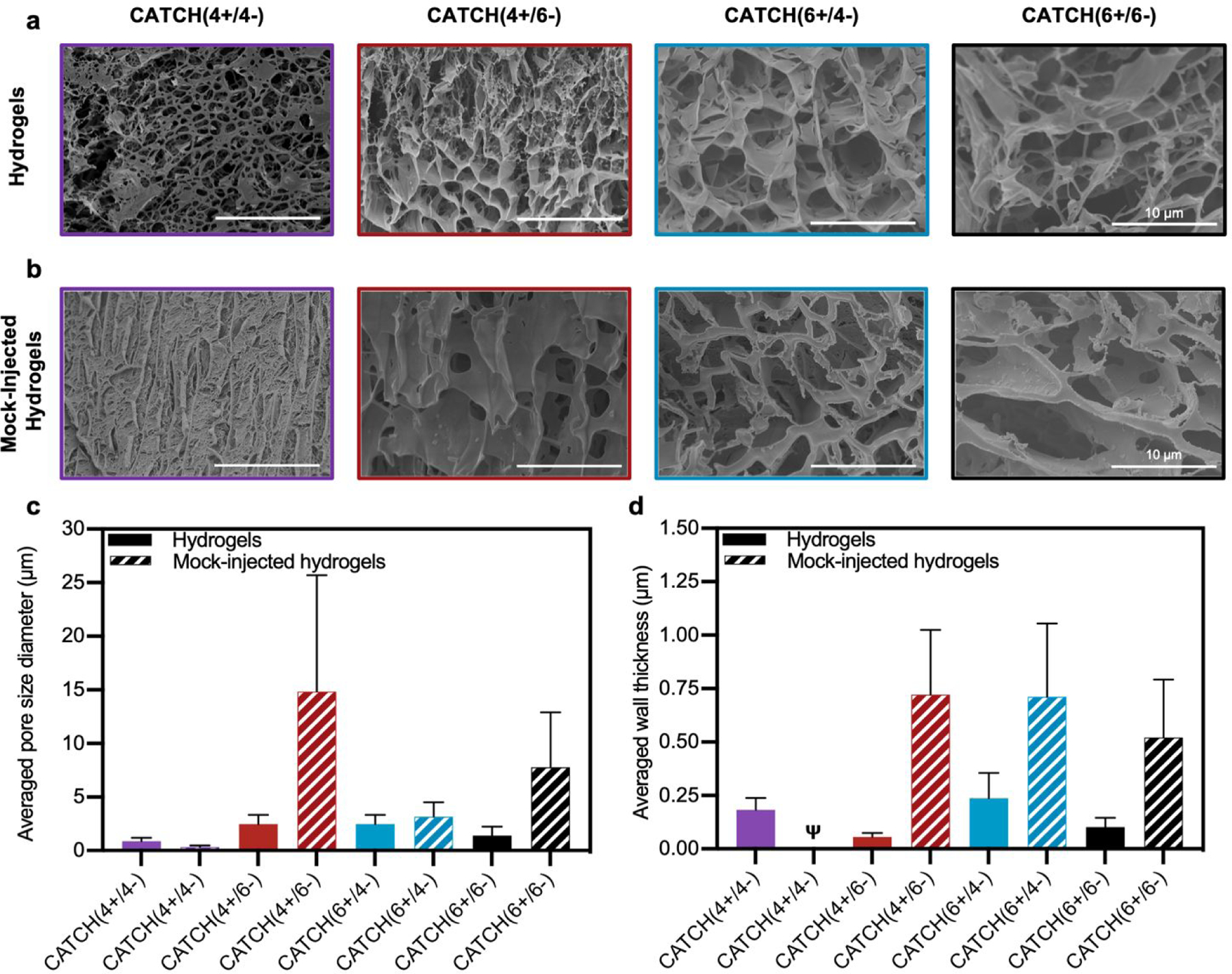
Figure 6. Cryogenic electron microscopy analysis of CATCH(+/−) hydrogel morphology.

(a) Cryo-SEM micrographs of 12 mM CATCH(4+/4−) CATCH(4+/6−), CATCH(6+/4−), and CATCH(6+/6−) hydrogels in the hydrated state. (b) Cryo-SEM micrographs of 12 mM CATCH(4+/4−), CATCH(4+/6−), CATCH(6+/4−), and CATCH(6+/6−) mock-injected hydrogels. (c) Average pore diameter measured from Cryo-SEM micrographs of CATCH(+/−) hydrogels and mock-injected hydrogels (Figure 5a–b) (n = 30 measurements). (d) Average wall thickness measured from Cryo-SEM micrographs of CATCH(+/−) hydrogels and mock-injected hydrogels (Figure 5a–b) (n = 30 measurements). Ψ indicates that wall thickness could not be reliably quantified from the Cryo-SEM micrographs.
