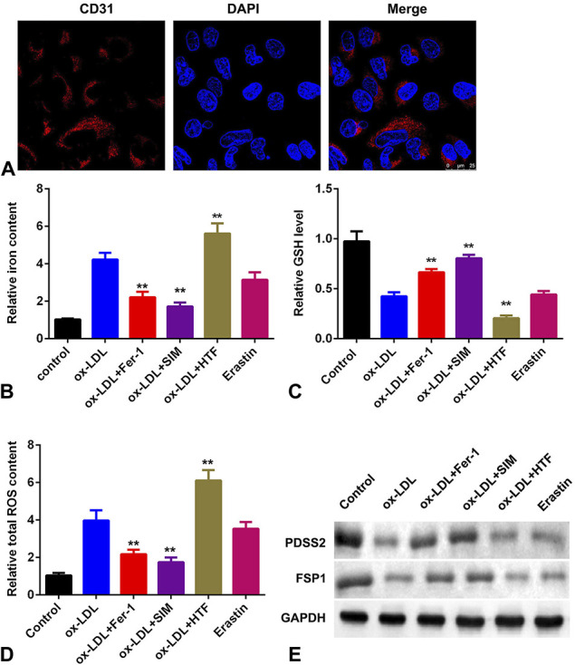
FIGURE 2.

Ox-LDL promotes the ferroptosis of HCAECs. A, Mitochondrial damage detected by immunofluorescence assay. B, The level of iron content in HCAECs. C, The levels of GSH. D, The total content of ROS. E, The protein levels of PDSS2 and FSP1. n = 3. **P < 0.01.
