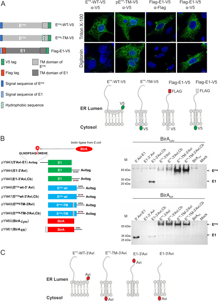
FIG 6.
Membrane topology of E1. (A) The upper part shows a schematic representation of constructs used in an immunofluorescence analysis: Erns wild type with C-terminal V5 tag, Erns TM with leucine stretch instead of the amphipathic helix and C-terminal V5 tag (18), and double-tagged E1 variant with an N-terminal Flag tag and a C-terminal V5 tag. On the right side, the results of immunofluorescence analyses are shown. BHK-21 cells were transfected with the indicated plasmids and fixed with 4% PFA on the following day. Cell membranes were permeabilized with Triton X-100 (all membranes) or the plasma membrane was selectively permeabilized with digitonin, followed by staining with specific antibody combinations: Erns, α-V5, α-mouse Alexa-Fluor-488; E1, α-FLAG, α-mouse Alexa-Fluor-488 or α-V5, and α-mouse Alexa Fluor 488; and nucleus, DAPI (blue). Below the micrographs are schematic representations of the membrane topology of the analyzed proteins. (B) As a second approach, in vivo labeling of proteins via BirA-mediated biotinylation of Avi tag-labeled proteins was conducted. On the left are constructs used in this section. The Avi-tagged proteins were transiently expressed in RK-13 cells cotransfected with pYM-48 (BirA cytosol, upper right) or pYM-49 (BirA ER, bottom right), and the expression products were analyzed for biotinylated proteins via Western analyses with α-streptavidin PO. (C) Schematic representation of membrane topology of Avi-tagged constructs.

