FIG 2.
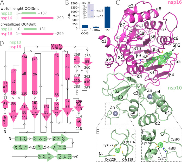
The crystal structure of the methyltransferase complex (nsp10:nsp16) from OC43-CoV. (A) Schematic representation of the crystallized and full-length nsp10:nsp16 proteins. (B) SDS-PAGE gel illustrating the purity of the recombinant nsp10:nsp16 complex and a graph illustrating its enzymatic activity. Values are for the reaction performed with all components except the enzyme (1st bar), with all components except for the RNA (2nd bar), and with all components (3rd bar). Error bars show standard deviations. A.U., arbitrary units. (C) Overall crystal structure of the nsp10:nsp16 complex (ribbon) with a sinefungin molecule (white) bound in the active site (SFG) and a “kick-out” omit map (Fo − Fc) of electron density (green) at 3.5 sigma with SFG excluded from the calculation. (D) Topological representation of the secondary-structure features of the complex. (E and F) Zn2+ coordination centers in nsp10 in kick-out omit maps (Fo − Fc) of electron density (green) at 10 sigma with Zn2+ excluded from the calculation.

