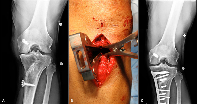
Figure 4.
Medial opening wedge high tibial osteotomy. A, Preoperative radiographs of a 45-year-old individual with symptomatic medial compartment OA. B, Lamina spreaders are used to gently distract the osteotomy once complete. C, Two-year follow-up PA flexion weight-bearing radiographs showing preserved medial compartment joint space with mild OA progression. OA = osteoarthritis

