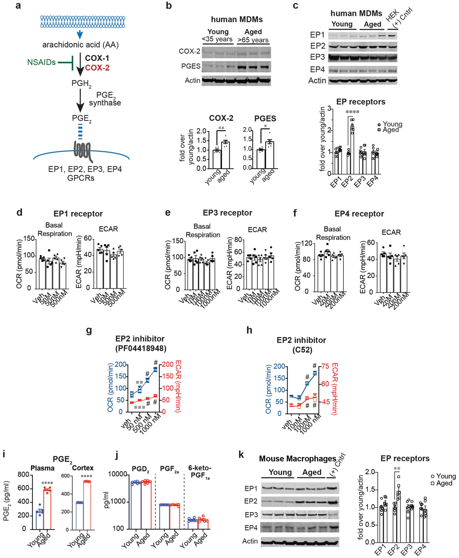
Extended Data Fig. 1 |. PGE2 regulates macrophage energy metabolism via the EP2 receptor.

Data are mean ± s.e.m. unless otherwise specified. a, The PGE2 synthetic pathway. Arachidonic acid is converted to the prostaglandin precursor PGH2 by the action of constitutive COX-1 and/or inducible COX-2; this step is inhibited by non-steroidal anti-inflammatory drugs (NSAIDs). PGE2 synthase converts PGH2 to PGE2 which can bind to four distinct G-protein-coupled receptors (EP1–EP4 receptors). b, Top, representative immunoblot of two independent experiments quantifying COX-2 and PGE2 synthase in young and aged human MDMs. Bottom, quantification shows higher COX-2 and mPGE2 synthase levels in aged (above 65 years) compared to young (below 35 years) human MDMs (n = 6 donors per group; **P = 0.0030, *P = 0.0106, Student’s two tailed t-test). c, Top, representative immunoblot of two independent experiments quantifying EP1–EP4 in young and aged human MDMs; last lane, HEK cells transfected with EP receptor cDNA as a positive control. Bottom, quantification shows increased EP2 in aged (over 65 years) compared to young (below 35 years) human MDMs (n = 6 donors per group; ****P < 0.0001, Student’s two tailed t-test). d–f, Quantification of basal respiration and ECAR of human MDMs treated with ascending doses of the EP1 agonist iloprost (d), the EP3 agonist AE248 (e) and the EP4 agonist AE1329 (f) for 20 h (n = 5 donors per group). g, h, Dose response of the brain-impermeant EP2 antagonist PF-04418948 (g) and the brain-penetrant EP2 antagonist C52 (h) in human MDMs at 20 h. Data are represented as box plots (5–95 percentile); one-way ANOVA for OCR and ECAR, P < 0.0001; Tukey’s post hoc test for PF-04418948: **P = 0.0083, ***P = 0.0005, #P < 0.0001; Tukey’s post hoc test for C52: #P < 0.0001 (n = 5 donors per group). i, LC–MS quantification of PGE2 levels in the plasma and cerebral cortex of aged (18–20 months) and young (3–4 months) mice. ****P < 0.0001 by two-tailed Student’s t-test (n = 6 mice per group) j, LC–MS quantification of PGD2, PGF2a and prostacyclin (measured as 6-keto-PGF1a) in the cerebral cortex of young and aged mice (n = 5–6 mice per group). k, Left, Representative immunoblot of EP1–EP4 levels in mouse peritoneal macrophages isolated from young (3–4 months) and aged (18–20 months) mice; last lane, HEK cells transfected with EP receptor cDNAs as a positive control (n = 6 mice per group). Right, EP2 is selectively upregulated in aged mouse peritoneal macrophages, similar to aged human MDMs in c; **P = 0.0018 (n = 6 mice per group).
