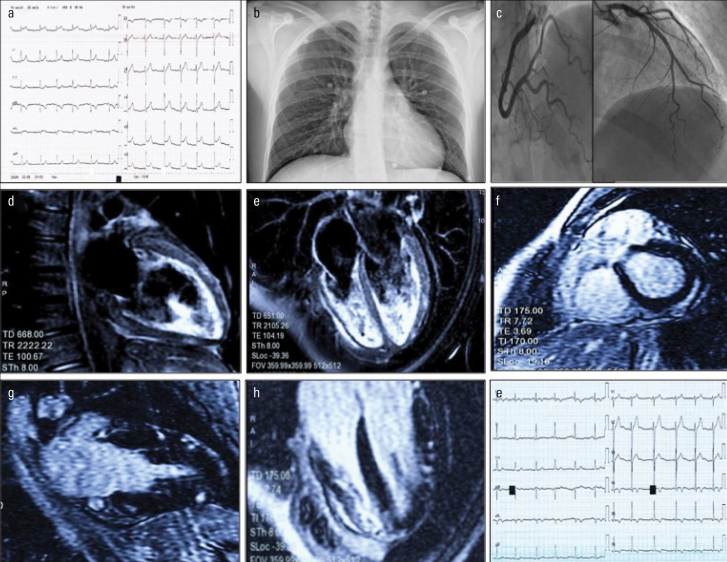
Figure 1.
a: Diffuse concave ST elevation with slightly widened QRS (90 ms, Axis + 60°)
b: Normal pulmonary texture on chest X-ray
c: Coronary arteries free from stenosis, TIMI 3 flow on all vessels
d and e: Patchy myocardial edema (water-sensitive T2-weighted sequence) in the anterior, inferior, and lateral walls. Mild pericardial effusion
f, g and h: Patchy epicardial hyperenhancement (late gadolinium enhancement sequence) in the posterior, anterior, inferior, and lateral walls
i: Electrocardiogram after one week with ST/T changes (note the negative T in the inferolateral site consensual to the signal changes reported on cardiac magnetic resonance)

