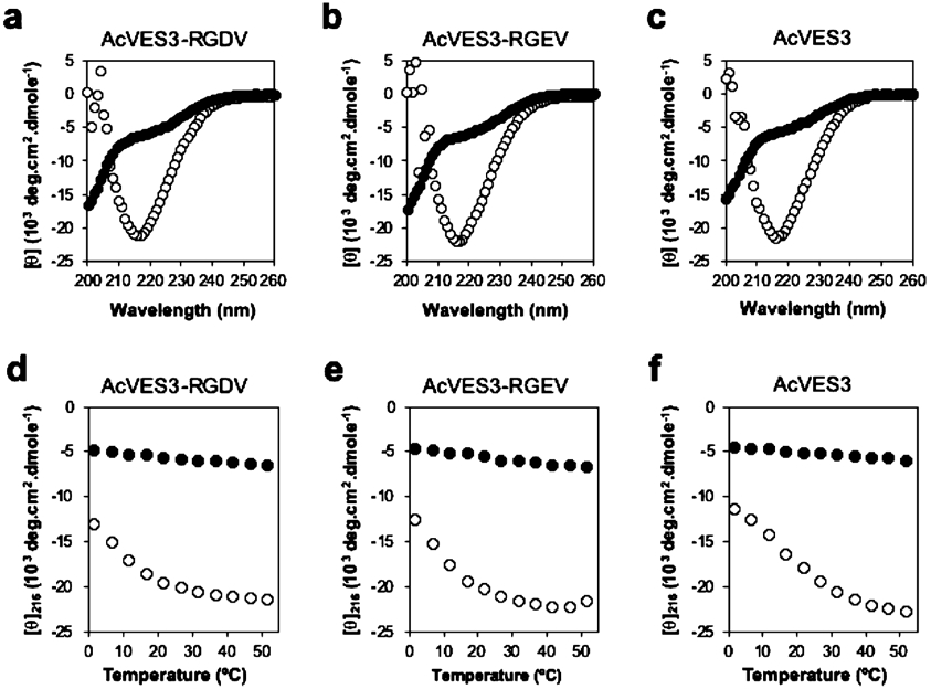
Figure 2.

(a–c) CD wavelength spectra of 150 μM peptides in water (●) or in HEPES buffer (○) (25 mM HEPES, 150 mM NaCl, pH 7.4) at 37 °C. (d–f) Temperature-dependent CD following [θ]216 in HEPES buffer. Increased signal at 216 nm is indicative of β-sheet structure formation.
