
Figure 3.
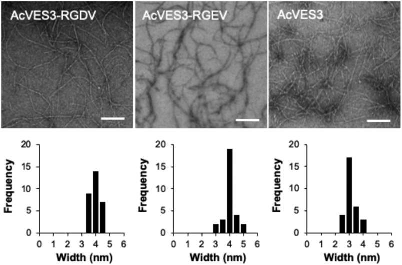
Transmission electron micrographs showing local morphology of fibrils formed by AcVES3-RGDV, AcVES3-RGEV, and AcVES3 peptides. The peptide gels (0.5 wt %) in HEPES buffer (25 mM HEPES containing 150 mM NaCl, pH 7.4) were formed by incubating overnight at 37 °C. Then, the TEM samples were prepared by suspending 5 μL of the resulting peptide gel in 195 μL of water to yield a 40-fold dilution. Fibril widths were determined from 30 independent measurements (scale bar = 100 nm).