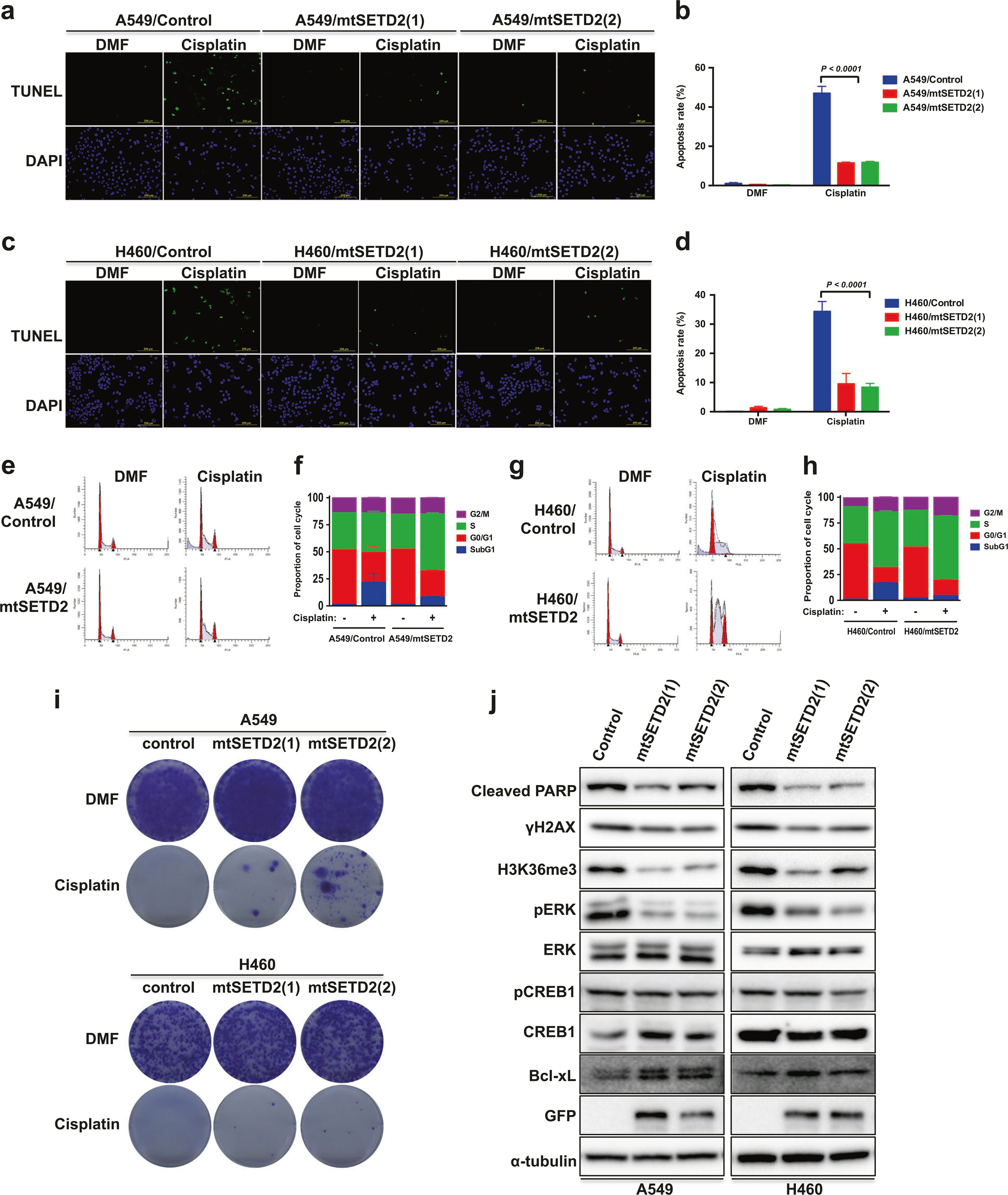
Fig. 5.

Ectopic expression of mutant SETD2 inhibits cisplatin-induced apoptosis in NSCLC cells. a Representative images of TUNEL staining of A549 or mtSETD2-expressing A549 cells after 15 μM cisplatin treatment for 24 h. A549 and H460 cells transfected with pBI-MCS-EGFP were used as control. b Representative histogram showing the percentage of apoptotic cells (TUNEL-positive cells). c Representative images of TUNEL staining of H460 or mtSETD2-expressing H460 cells after 15 μM cisplatin treatment for 24 h. d Representative histogram showing the percentage of apoptotic cells (TUNEL-positive cells). Data (b,d) are means ± s.d. from three independent experiments, and statistical significance was determined by two-way ANOVA with Sidak’s multiple comparisons test. e A549 or mtSETD2-expressing A549 cells were exposed to 15 μM cisplatin for 24 h and the cell cycle distribution was determined by flow cytometry analysis. f Representative cell cycle histogram of (e) demonstrating the percentage of the cells in each phase. Data are means ± s.d. from n = 3. g H460 or mtSETD2-expressing H460 cells were exposed to 15 μM cisplatin for 24 h and the cell cycle distribution was determined by flow cytometry analysis. h Representative cell cycle histogram of (g) demonstrating the percentage of the cells in each phase. Data are means ± s.d. from n = 3. i Cell survival of mtSETD2-expressing A549 or H460 cells under 10 μM cisplatin treatment for 24 h was assessed by clonogenic assay. j Mutant SETD2-expressing A549 or H460 cells were exposed to 10 μM cisplatin for 24 h, and WB analysis was performed
