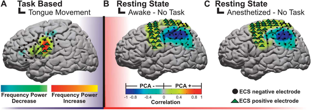
FIGURE 3.
Examples of electrocorticographic brain mapping. A, example of cortical topography of a gamma power alteration associated with a subject protruding his or her tongue. The positive motor stimulation sites are represented by green triangles. B, example of resting-state networks derived from a principal component analysis (PCA) while the patient is awake. C, example of resting-state networks derived from a PCA while the patient is under anesthesia. Notably, these networks are preserved regardless of level of consciousness and can be derived in the absence of task. ECS, electrocortical stimulation.

