Figure 2.
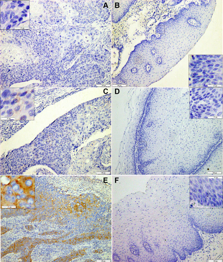
Immunohistochemical staining of PABPC1 in ESCC.
Notes: Representative images of PABPC1 staining with low expression (A, C) and high expression (E) in ESCC tissues and their paired adjacent normal epithelium tissues (B,D, F, respectively). Abbreviation: ESCC, esophageal squamous cell carcinoma.

