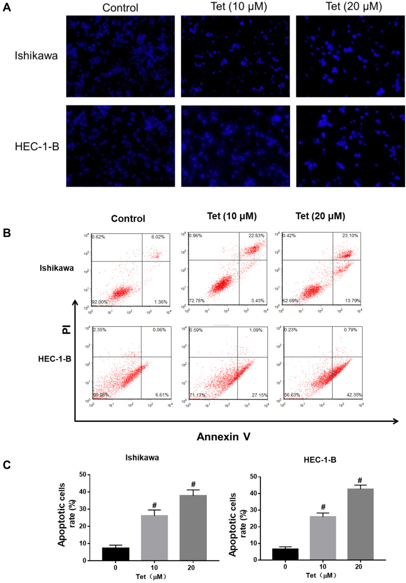
Figure 7.
Tetrandrine induced DNA fragmentation and apoptosis in endometrial cancer cells. The Ishikawa and HEC-1-B cells were treated with 0, 10 or 20µM tetrandrine for 24h. (A) Hoechst 33258 staining showed typical apoptotic morphology changes (scale bars: 100 μm). (B) The cell apoptosis was assessed by Annexin V- FITC/PI staining. (C) Histogram analysis showed the percentage of apoptotic. Data were presented as means ± SD (n=3), #P<0.05.

