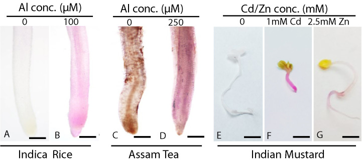
Figure 2. Histochemical determination of lipid peroxidation in different plant species by Schiff’s reagent.
Detection of lipid peroxidation using Schiff’s reagents in Indica rice root (A) control, scale bar = 100 μm; (B) stressed, scale bar = 100 μm; Assam tea roots (C) control, scale bar = 100 μm, (D) stressed, scale bar = 100 μm; and in Indian mustard seedling (E) control, scale bar = 5 mm, (F) 1 mM Cd treated seedling, scale bar = 5 mm, (G) 2.5 mM Zn treated seedling, scale bar = 5 mm.

