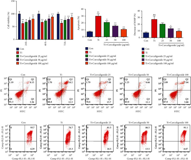
Figure 1.

Curculigoside treatment attenuated Ti-induced inhibition of osteoblastic differentiation in MC3T3-E1 cells. (a) After being intervened with Ti and treated with curculigoside at different concentrations (25, 50, and 100 μg/ml) for 24 h, 48 h, and 72 h, MC3T3-E1 cell viability was detected by the CCK-8 assay (n = 6). (b, d) After being intervened with Ti and treated with curculigoside, cell apoptosis was detected by flow cytometry (n = 3). (c, e) Mitochondrial membrane potential of MC3T3-E1 cells cultured under Ti and different concentrations of curculigoside was assayed by flow cytometry using JC-1 assay kits, and the decrease in the MMP index in each group cell was quantified (n = 3). Results are represented as the mean ± SD. ▲P < 0.05, compared to the Con group; ▲▲P < 0.01, compared to the Con group; ∗P < 0.05, compared to the Ti group; ∗∗P < 0.01, compared to the Ti group. Ti: titanium particle; Con: control; MMP: mitochondrial membrane potential.
