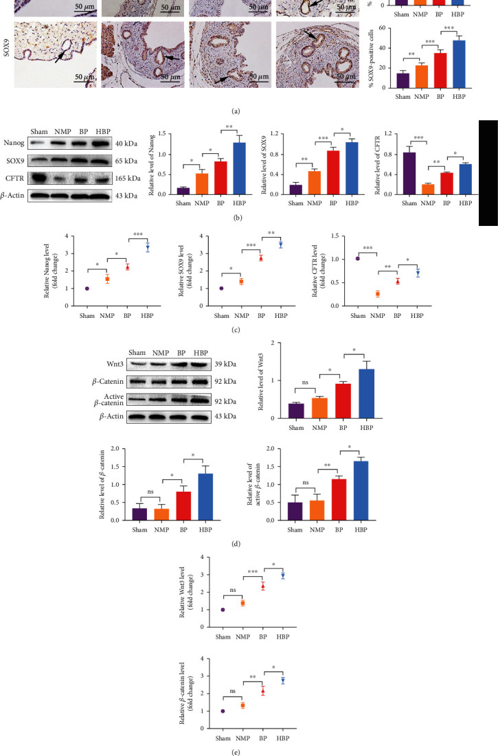
Figure 5.

Increased stemness and activation of the Wnt signal pathway in PBGs. (a) Pluripotent cell marker- (Nanog- and SOX9-) positive cells in PBGs were marked by immunohistochemistry (arrow). Nanog- and SOX9-positive cells in the sham group were 15.10% ± 2.19 and 14.90% ± 2.56, respectively, slightly increased in the NMP group (35.00 ± 5.78%, P < 0.0001 vs. the sham group, 22.70 ± 2.44%, P = 0.0097 vs. the sham group) and significantly increased in the BP group (45.20 ± 4.44%, P = 0.00123 vs. the NMP group and 35.10 ± 3.32%, P = 0.0001 vs. the NMP group, respectively) and the HBP group (58.40 ± 4.88%, P = 0.0015 vs. the BP group and 47.70 ± 4.60%, P = 0.0001 vs. the BP group). Relative levels of Nanog, SOX9, and CFTR in each group were detected by western blotting (b) and PCR (c) (n = 3). (d) Western blotting analysis of Wnt3, total β-cat, and active-β-cat showed no statistical differences between the NMP group and the sham group, while expression of Wnt3, total β-cat, and active-β-cat increased in the BP and HBP groups (n = 3). (e) Relative levels of Wnt3 and Ctnnb1 (total β-cat) levels were detected using PCR (n = 3). ∗P < 0.05, ∗∗P < 0.01, and ∗∗∗P < 0.001. PBG: peribiliary gland; NMP: normothermic machine perfusion; HBP: HO-1/BMMSCs plus NMP; BP: BMMSCs plus NMP; SOX9: SRY-box transcription factor 9; CFTR: cystic fibrosis transmembrane conductance regulator; β-cat: β-catenin.
