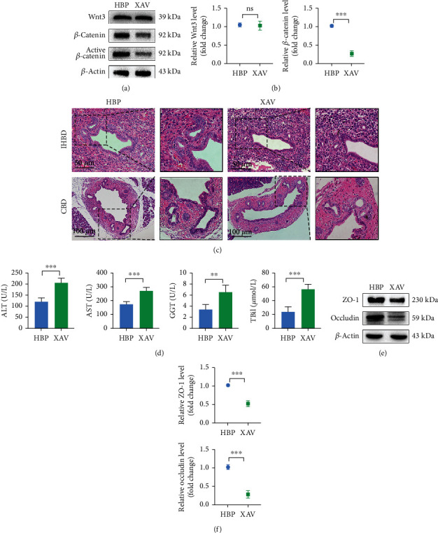
Figure 6.

Wnt signal pathway triggered proliferation of biliary progenitor cells to repair bile duct injury. Activation levels of the Wnt signal pathway in each group were detected by western blotting (a) and PCR (b) (n = 3). (c) Histological changes of bile ducts and PBGs after administration of XAV-939. (d) Liver function changes after administration of XAV-939. ALT, AST, GGT, and TBil in the HBP group were 120.70 ± 17.96 U/L, 176.40 ± 18.61 U/L, 24.12 ± 7.26 U/L, and 3.42 ± 0.93 μmol/L, respectively, while they were worse in the XAV-939 group (207.40 ± 20.41 U/L, 273.8 ± 24.11 U/L, 56.92 ± 7.32 U/L, and 6.58 ± 1.26 μmol/L, respectively). Relative levels of tight junctions (ZO-1 and occludin) in each group were detected using western blotting (e) and PCR (f) (n = 3). ∗P < 0.05, ∗∗P < 0.01, and ∗∗∗P < 0.001. PBG: peribiliary gland; ALT: alanine aminotransferase; AST: aspartate aminotransferase; GGT: gamma-glutamyl transpeptidase; TBil: total bilirubin; HBP: HO-1/BMMSCs plus NMP.
