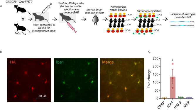
Figure 1.
Isolation and validation of microglia-specific mRNA from Cx3cr1CreER: Rpl22HA mice. (A) Overview of the experimental strategy of generation of Cx3cr1CreER: Rpl22HA mice and isolating RNA. Black ribosomes in the cartoon represent endogenous wt ribosomes, whereas yellow/red are HA tagged transgenic Rpl22 containing ribosomes. (B) Representative images from the amygdala of Cx3cr1CreER: Rpl22HA mice validating the specificity of HA expression in microglia. CX3CR1 + cells carry GFP and an antibody against GFP was used to label CX3CR1 + cells (green) and anti-HA was used to label Rpl22HA expressing cells. The merged imaged shows that HA and GFP are co-localized. (C) Bar graph shows that RNA is significantly enriched in the immunoprecipitated RNA. *indicates p < 0.05 (Tukey’s multiple comparison test). The enrichment in the microglial IP RNA was compared to the total RNA before IP. Three animals per group were used in the experiments described in (B,C).

