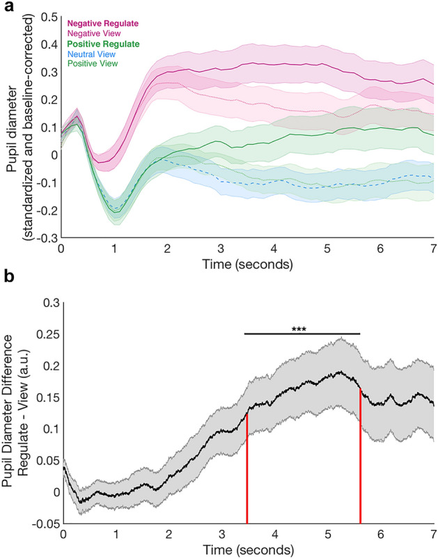
Figure 2.

Pupil results. (a) Mean pupil dilation during the 7-s regulate (saturated colours) and view periods (lighter colours). The signal across the whole emotion task was z-scored within-participant and baseline-corrected on each trial for the pupil size in the 500 ms prior to the regulation period (during which the phase-scrambled adaptation version of the stimulus was displayed, see “Materials and methods”). The mean pupil dilation was calculated over all 20 trials in each block for each participant and then averaged across the group. The shaded areas indicate the standard error of the mean across participants. Lines denote the mean pupil dilation at each time point. The dotted blue line represents the neutral view condition for comparison. (b) Pupil Diameter Difference between Regulation and View Trials. We collapsed over positive and negative blocks in order to test for a valence-independent regulation signal across all participants. The mean z-scored pupil dilation for positive and negative view trials was subtracted from the mean z-scored dilation during positive and negative reappraise trials for each participant. A cluster-based permutation test indicated that between 3.4 and 5.6 s of the reappraisal/view period, the pupil dilation in reappraisal blocks was larger than during viewing blocks (p < 0.001; indicated by the black horizontal line and stars). For each participant, the mean of the Reappraisal – View difference in the pupil dilation signal during this period (marked by the red vertical lines in the plot) determines the Pupil Dilation Index. The shaded areas indicate the standard error of the mean across participants. The black graph denotes the mean pupil dilation at each time point.
