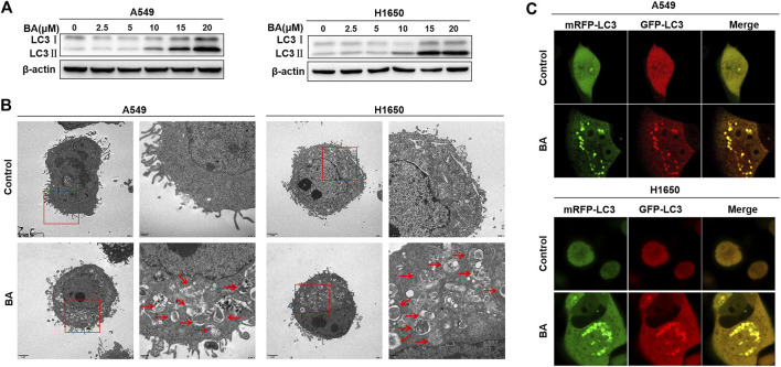
FIGURE 2.
BA induces autophagy in NSCLC cells. (A) A549 and H1650 cells were treated with the indicated concentrations of BA for 48 h, and the expression of autophagy-related protein LC3 was analyzed by Western blots. (B) A549 and H1650 cells were treated with 15 μM BA for 48 h, and the autophagic vacuoles were observed using transmission electron microscopy. (C) A549 and H1650 cells were transfected with adenovirus expressing mRFP-GFP-LC3, then treated with 15 μM BA for 48 h, and representative images were obtained using a confocal microscope.

