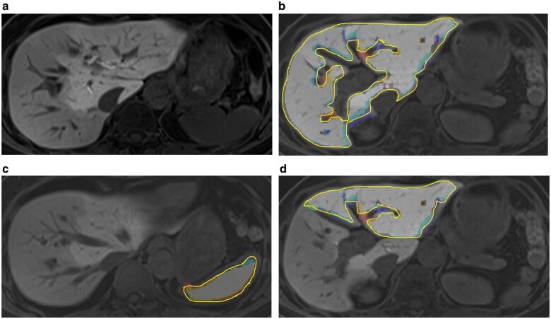
Fig. 1.
Example of gadoxetate disodium-enhanced MRI and segmentation in a patient with hilar cholangiocarcinoma located in the right hepatic duct with unilateral biliary drainage of the left liver and preoperative portal vein embolization
a Original gadoxetate disodium-enhanced MRI (EOB-MRI)); b segmented whole liver; c segmented spleen; and d segmented future remnant liver (FLR) after right hemihepatectomy.

