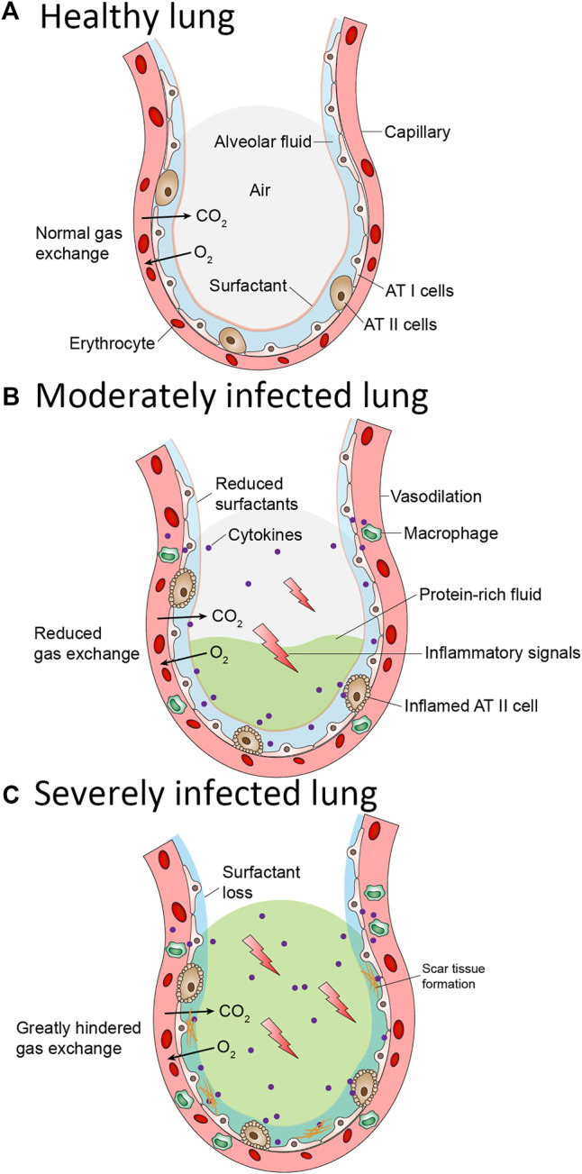
FIGURE 2.

Pathological changes in the lung alveolus during COVID-19 (A) Normal alveolus is wrapped with capillaries containing red blood cells. Oxygen in the alveolus is exchanged with carbon dioxide in the capillaries. The alveolus surface contains alveolar Type I and Type II cells. Type I cells enables gas exchange. Type II cells secrete pulmonary surfactant (PS). PS lines the alveolus and prevent it from collapsing (B) In a moderately infected lung, Alveolar Type II cells are inflamed resulting in reduced pulmonary surfactant. Surface tension and pressure increase inside the alveolus affecting the gas exchange. Vasodilation of the capillary occurs resulting in the release of inflammatory cytokines and accumulation of protein-rich fluid inside the alveolus (C) In severely infected lung, the alveolar type II cells become more inflamed thereby resulting in complete loss of pulmonary surfactant. Scar tissue on the alveolar surface began to form. The release of inflammatory cytokines is increased, and more protein-rich fluid accumulate inside the alveolus. The oxygen/carbon dioxide exchange is greatly hindered and thus patients in this stage must undergo intubation as an aid to breathe.
