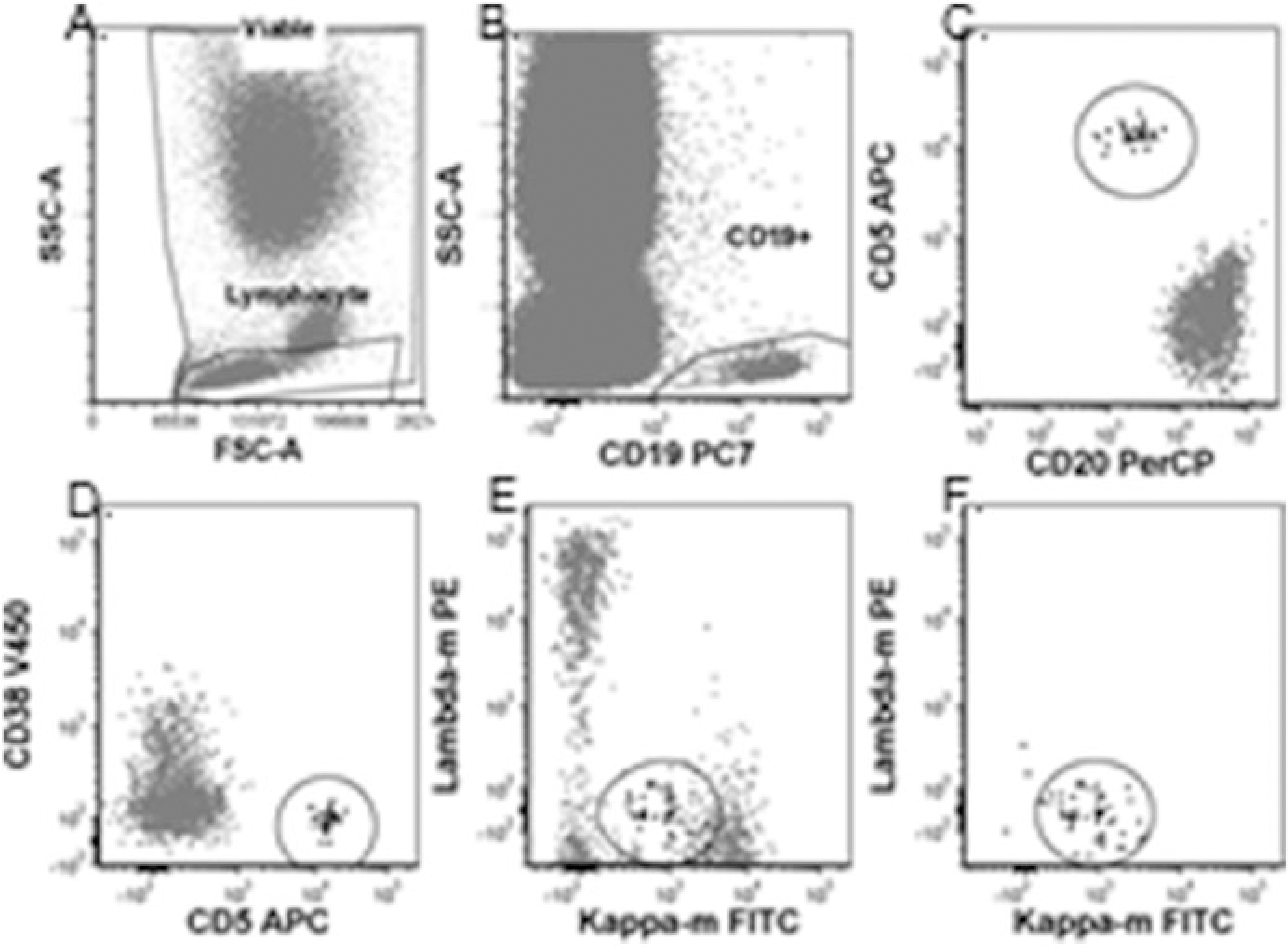
Fig. 4.

CLL MRD analysis method 2: (a) Viable and lymphocyte gates. (b) CD19+ gate. (c) Gated on viable, singlet, lymphocyte, and CD19+ gates. Abnormal CLL cells CD20 dim and CD5+ in ellipse. (d) Gated on viable, singlet, lymphocyte and CD19+ gates. Abnormal CLL cells CD5+ and homogeneously CD38 negative in ellipse. (e) Gated on viable, singlet, lymphocyte and CD19+ gates. Abnormal CLL cells in ellipse masked by polyclonal B cells. (f) Gated on viable, singlet, lymphocyte, CD19+, and abnormal CD5, CD20, and CD38 gates. Abnormal CLL cells in ellipse are monoclonal, dim positive for kappa and negative for lambda
