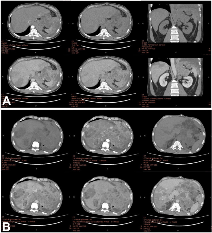
Figure 1.
(A) CT scan with four-phase contrast shows an irregular infiltrative heterogeneous enhanced mass involving the left hemidiaphragm and left adrenal gland mass with several hypodense lesions scattered throughout the lobes of the liver. No radiological evidence supports liver cirrhosis. (B) The CT study shows an interval increase in the left adrenal gland mass, now measuring about 11.2×15.2×5.5 cm. Interval increased extension of necrotic soft tissue and multiple matted necrotic nodes involving the left hemidiaphragm, pericardial fat pad, pericardium, periaortic, gastrohepatic and peripancreatic regions, which are unprecedented. This lesion shows the direct invasion of the distal oesophagus and gastric cardia, causing proximal oesophageal dilation. Increased extension of tumour thrombus in inferior vena cava extending into the right hepatic vein ascends to the right atrium and extension in the left inferior pulmonary vein reaching into the left atrium. These conditions progress within 3 weeks after the first CT study.

