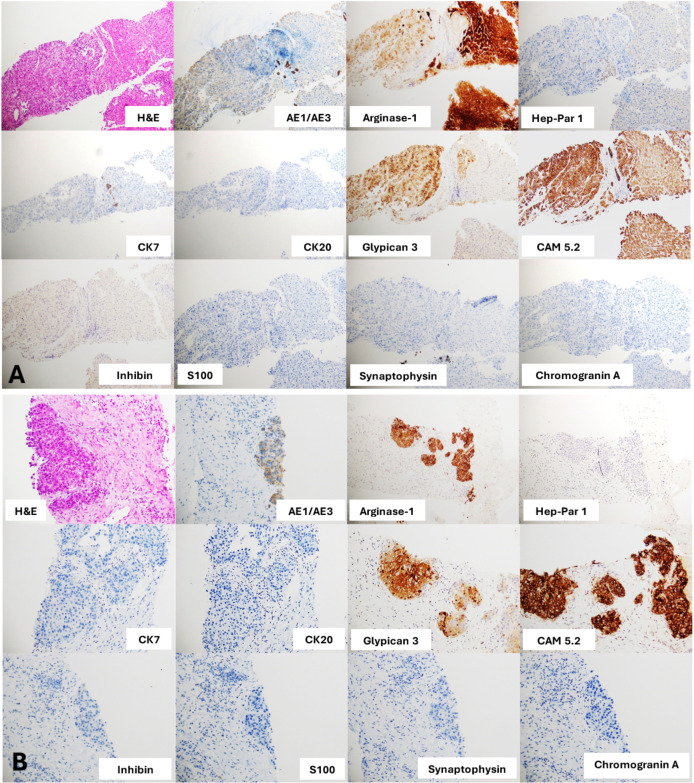
Figure 2.
(A) Sections of liver nodule core biopsies show an epithelial neoplasm consisting of the proliferated polygonal epithelial cells arranged in 6–7 cell thick trabeculae and nested separately by flat endothelial lining sinusoidal spaces. The neoplastic cells contain vesicular and slightly pleomorphic irregular thick nuclear membrane nuclei, prominent nucleoli and rare mitoses. Intranuclear cytoplasmic inclusions are noted. Adjacent normal liver parenchyma is present on the right side of the picture. These biopsies appear to have positive stain for AE1/AE3, arginase-1, glypican-3 and CAM5.2, but a negative stain for CK7, CK20, inhibin, S100, synaptophysin or chromogranin A. Hepatocellular carcinoma or hapatoid adenocarcinoma is suggested from these results. (B) Sections of adrenal mass core biopsies show an epithelial neoplasm within a desmoplastic stroma. The adrenal mass shares similar histological features to the liver nodule; positive stain for AE1/AE3, arginase-1, glypican-3 and CAM5.2 and negative stain for CK7, CK20, inhibin, S100, synaptophysin or chromogranin A. No residual non-neoplastic adrenal tissue was present in the core biopsies. Thus, we can conclude the resemblance of both liver nodules and adrenal mass.

