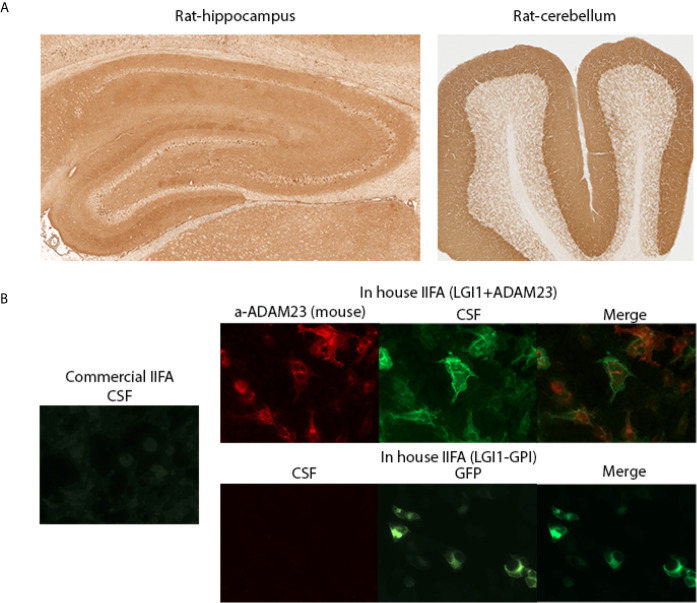
Figure 3.
Discrepancies identifying LGI1 antibodies by IIFA. (A) A patient’s CSF demonstrating LGI1 immunoreactivity on rat brain immunohistochemistry. Hippocampus (left panel) and cerebellum (right panel) staining patterns. (B) Left panel shows patient’s CSF reactivity on commercial IIFA LGI1 transfected cells (negative staining); Right panels show CSF reactivity on in-house IIFAs using LGI1 plus ADAM23 transfected cells (upper panels; positive staining) and GPI-LGI1 transfected cells (lower panels, negative staining). GPI: glycosyl-phosphatidylinositol anchored LGI1 (to display the protein on the cell surface).

