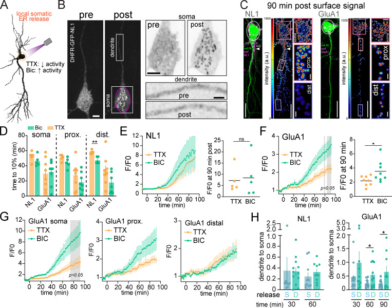
Figure 6.
Local release from the cell body ER reveals direct, long range trafficking to dendrites. (A) Schematic of experimental strategy. DHFR-GFP-NL1 and DHFR-GFP-GluA1 were locally released from the soma in the presence of TTX to suppress or Bic to elevate neuronal activity. (B) Example of DHFR-GFP-NL1 intracellular localization before (left) and 14 min after (right) somatic ER release (pink circle). The magnified images to the right show the soma (top) and a section of dendrite (bottom) before and after ER release. Note the absence of vesicular structures appearing in the dendrites shortly following somatic release. Scale bar, 6 µm. Top inset scale bar, 6 µm. Bottom inset scale bar, 3 µm. (C) Merged confocal images showing cell fill (green) and surface signal (Alexa647-anti-GFP, magenta) for DHFR-GFP-NL1 (left) and DHFR-GFP-GluA1 (right) 90 min following local ER release (white circles). Surface signal is shown in the heatmap images. Insets show the soma, proximal, and distal dendrites 90 min after release. Scale bars, 20 µm. Soma inset scale bars, 5 µm. Dendrite inset scale bars, 2 µm. (D) Time to 10% surface accumulation following somatic ER release in the presence of TTX (orange bars) or Bic (green bars) for NL1 and GluA1. Mean ± SEM; **, P < 0.01 (Student's t test; n = 5–8 neurons from at least three independent experiments). (E) Time course of DHFR-GFP-NL1 surface trafficking (over the entire cell) following somatic ER release in the presence of TTX (orange line) or Bic (green line). Surface signal at 90 min is shown to the right; mean ± SEM (Student's t test; n = 5 neurons/condition from three independent experiments). (F) Time course of DHFR-GFP-GluA1 surface trafficking (over the entire cell) following somatic ER release in the presence of TTX (orange line) or Bic (green line). The gray shaded regions designate P < 0.05, two-way ANOVA, Bonferroni’s multiple comparisons test; n = 8 neurons/condition from two independent experiments. Surface signal at 90 min is shown on the right; mean ± SEM; *, P < 0.05 (Student's t test). (G) Time course of DHFR-GFP-GluA1 surface trafficking at the soma (left), proximal dendrites (<40 µm from the soma; center), and distal dendrites (40–200 µm from the soma; right) in the presence of TTX or Bic; mean ± SEM. The gray shaded region designates P < 0.05, two-way ANOVA, Bonferroni’s multiple comparisons test. (H) The ratio of total dendritic to somatic signal is plotted at different time points following local ER release from the soma (blue) or from dendrites (teal) for DHFR-GFP-NL1 (left) and DHFR-GFP-GluA1 (right); mean ± SEM; *, P < 0.05 (Student's t test; n = 9–16 neurons from at least two independent experiments). dist., distal; prox., proximal.

