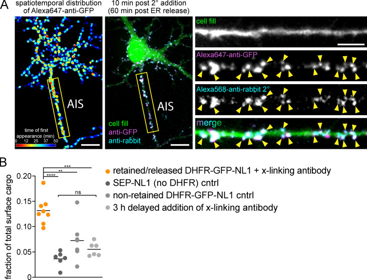
Figure S3.
Control experiments for surface NL1 accumulation at the AIS. (A) Antibody-labeled DHFR-GFP-NL1 signal is localized to the cell surface. DHFR-GFP-NL1 was released from the ER and continuously cross-linked and labeled with Alexa647-anti-GFP as it appeared at the cell surface for 50 min after ER release. Left: The timing and distribution of accumulated Alexa647-anti-GFP (generated in rabbit) immediately before addition of Alexa568-anti-rabbit secondary to label cell surface Alexa647-anti-GFP. The center panel shows the same neuron 10 min following addition of Alexa568-anti-rabbit (cyan). Insets to the right show the AIS, taken from the yellow box in the image to the left. The robust colocalization of Alexa647-anti-GFP and Alexa568-anti-rabbit (arrowheads) confirms accumulated DHFR-GFP-NL1 is on the cell surface. Scale bars, 10 µm. Inset scale bar, 5 µm. (B) Comparison of the fraction of total surface cargo at the AIS for retained/released DHFR-GFP-NL1 in the continuous presence of cross-linking antibody (orange) versus three trafficking controls: SEP-GluA1 (dark gray), nonretained (no zapalog) DHFR-GFP-NL1 (gray), and DHFR-GFP-NL1 that was released and allowed to traffic for 3 h before addition of cross-linking antibody (light gray). The retained/released DHFR-GFP-NL1 is the same data shown in Fig. 4 B with Alexa647-anti-GFP present for the duration of the experiment; mean ± SEM; **, P < 0.01; ***, P < 0.001; ****, P < 0.0001 (one-way ANOVA, Tukey’s multiple comparisons test; n = 8 [retained/released DHFR-GFP-NL1]; n = 6 [SEP-NL1]; n = 7 [nonretained DHFR-GFP-NL1]; n = 6 [3 h delayed addition cntrl]; n = number of neurons). cntrl, control.

