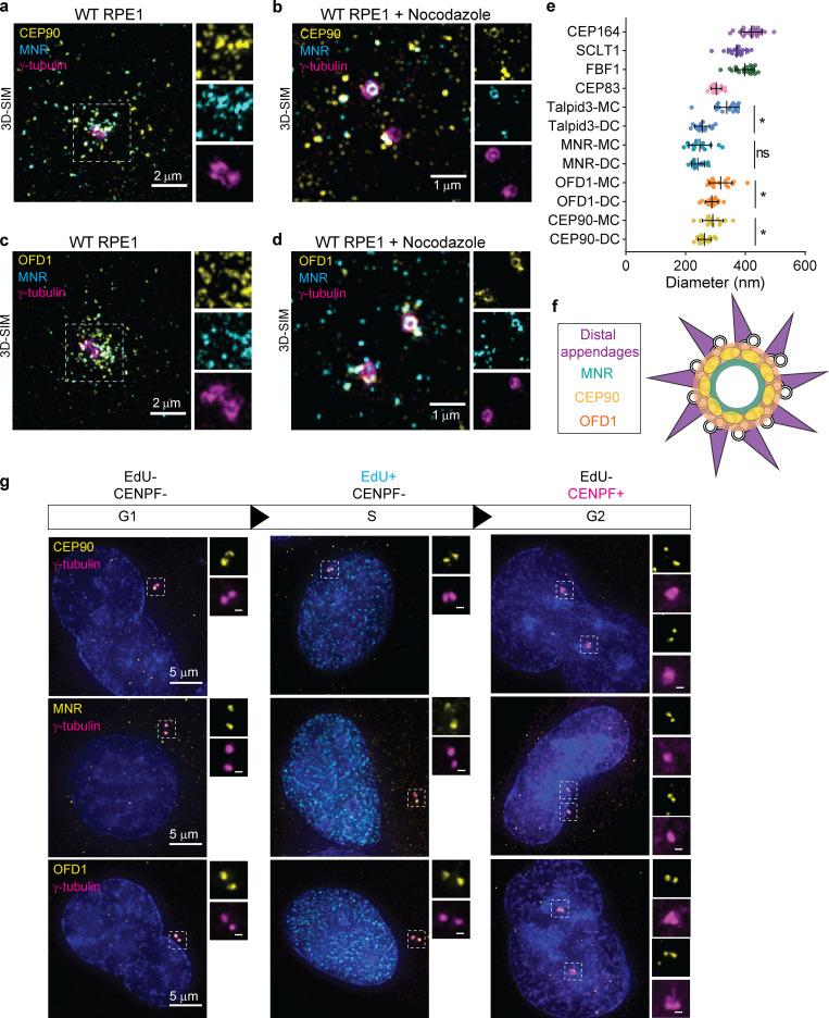
Figure 3.
CEP90 colocalizes with OFD1 and MNR. (a) Immunostaining of RPE1 cells for CEP90 (yellow), MNR (cyan), and γ-tubulin (magenta) demonstrating that CEP90 colocalizes with MNR at centriolar satellites. Scale bar = 2 µm. (b) 3D-SIM of RPE1 cells treated with nocodazole to disperse centriolar satellites highlights colocalization of CEP90 (yellow) and MNR (cyan) at centrioles (γ-tubulin, magenta). Scale bar = 1 µm. (c) Immunostaining of OFD1 (yellow), MNR (cyan), and γ-tubulin (magenta) reveals CEP90 and MNR colocalization at centriolar satellites. Scale bar = 2 µm. (d) 3D-SIM of immunostained cells treated with nocodazole reveals a ring of OFD1 (yellow) colocalizing with MNR (cyan) at centrioles (γ-tubulin, magenta). Scale bar = 1 µm. (e) Measurements of ring diameters measured in 3D-SIM images. For distal centriole proteins, ring diameters were measured at the mother (MC) and daughter centriole (DC). Scatter dot plot shows mean ± SD. *, P < 0.05, unpaired t test. n = 13–22 centrioles. (f) Schematic representation of a radial view of a mother centriole with distal appendages (purple), OFD1 (orange), CEP90 (yellow), and MNR (green). (g) Localization of distal centriole proteins (yellow) to centrioles (γ-tubulin, magenta) is cell cycle dependent. Cells in G1 phase were identified as lacking both EdU and CENPF staining, cells in S phase as being positive for EdU but lacking CENPF, and cells in G2 phase as being positive for CENPF but lacking EdU staining. Two puncta of CEP90, OFD1, and MNR were observed at one centrosome during G1 and S phases and four puncta at two centrosomes after centrosome duplication during G2 phase. Scale bars represent 5 µm in main panels and 0.5 µm in insets.

