Figure S5.
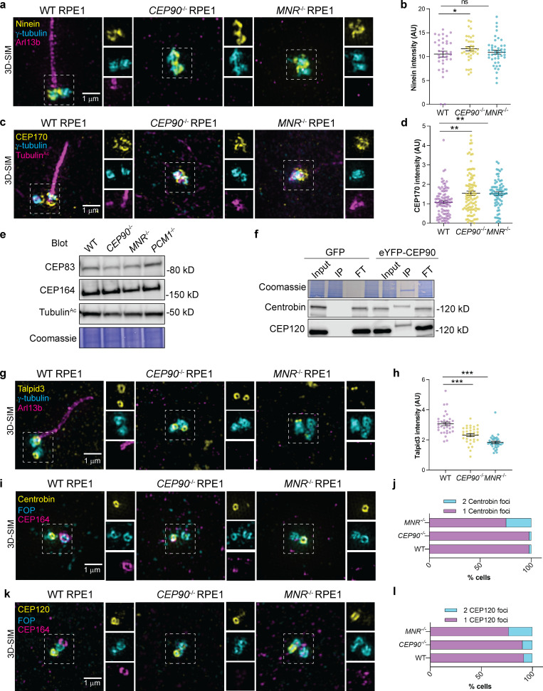
CEP90 regulates distal appendage assembly independent of Talpid3 recruitment and removal of Centrobin.(a) 3D-SIM imaging of serum-starved WT, CEP90−/−, and MNR−/− RPE1 cells immunostained for Ninein (yellow), centrioles (γ-tubulin, cyan), and cilia (ARL13B, magenta). Boxed regions are depicted in insets throughout. Ninein localizes to centrioles in WT, CEP90−/−, and MNR−/− cells. Scale bar = 1 µm. (b) Quantification of Ninein fluorescence intensity at centrioles in WT, CEP90−/−, and MNR−/− cells. Horizontal lines indicate means ± SEM. Asterisks indicate P < 0.05 determined using one-way ANOVA. n = 36–45 measurements. (c) 3D-SIM imaging of serum-starved WT, CEP90−/− and MNR−/− RPE1 cells immunostained for CEP170 (yellow), centrioles (γ-tubulin, cyan) and cilia (TubulinAc, magenta). Boxed regions are depicted in insets throughout. CEP170 localizes to centrioles in WT, CEP90−/−, and MNR−/− cells. Scale bar = 1 µm. (d) Quantification of CEP170 fluorescence intensity at centrioles in WT, CEP90−/−, and MNR−/− cells. Horizontal lines indicate means ± SEM. Asterisks indicate P < 0.005 determined using one-way ANOVA. n = 73–99 measurements. (e) Immunoblot of distal appendage proteins CEP83 and CEP164 in WT, CEP90−/−, MNR−/−, and PCM1−/− RPE1 cells. (f) Coimmunoprecipitation of daughter centriole proteins Centrobin and CEP120 with CEP90. IP indicates eluate, and FT indicates flow-through. (g) WT, CEP90−/−, and MNR−/− RPE1 cells were serum starved for 24 h and stained with antibodies to Talpid3, γ-tubulin (centrosome marker), and ARL13B (cilia marker). 3D-SIM imaging reveals ring of Talpid3 at mother and daughter centrioles. Scale bars represent 1 µm for main panels and insets. (h) Quantification of Talpid3 fluorescence intensity at centrioles. Scatter dot plots show mean ± SEM. Asterisks indicate P < 0.0005, determined using one-way ANOVA. n = 33–35 measurements. (i) WT, CEP90−/−, and MNR−/− serum-starved RPE1 cells immunostained for Centrobin (yellow), centrioles (FOP, cyan), and distal appendages (CEP164, magenta). Scale bar = 1 µm. (j) Quantification of whether Centrobin localizes to one or two centrioles. n > 50 cells from two biological replicates. CEP90 and MNR are not required to remove Centrobin from the distal mother centriole. (k) WT, CEP90−/−, and MNR−/− serum-starved RPE1 cells immunostained for CEP120 (yellow), centrioles (FOP, cyan), and distal appendages (CEP164, magenta). Scale bar = 1 µm. (l) Quantification of whether CEP120 localizes to one or two centrioles. n > 50 cells from two biological replicates. CEP90 and MNR are not required to remove Centrobin or CEP120 from the distal mother centriole.

