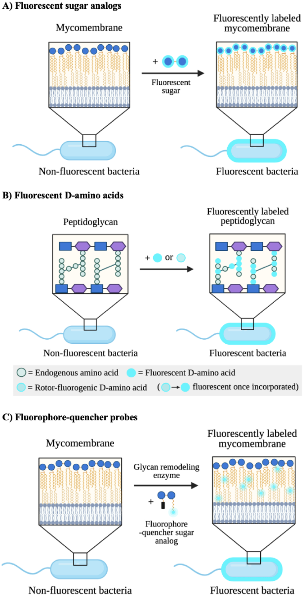
Figure 10. Mechanisms of action of metabolic imaging probes.

A) Fluorescent sugar analogs are metabolically incorporated into endogenous glycans to enable their detection and tracking via fluorescence. Trehalose is depicted by two linked blue circles. B) Fluorescent and rotor-fluorogenic D-amino acids are metabolically incorporated into peptidoglycan and facilitate the real-time imaging of peptidoglycan biosynthesis. Rotor-fluorogenic D-amino acids turn fluorescent once incorporated into cellular peptidoglycan. C) Fluorophore-quencher probes accessed from sugar analogs become fluorescent upon specific enzyme-catalyzed reactions and allow the direct visualization of enzyme activity during cell wall biosynthesis.
