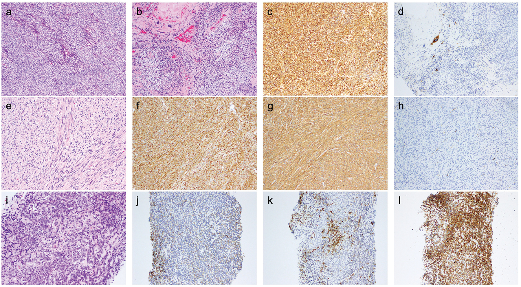
Figure 3. Histopathologic features of selected cases (see text and tables for further details).

a-d) UMT04 - myogenic sarcoma, most likely leiomyosarcoma. a and b) H&E – tumor composed of tight fascicles of spindled cells with fusiform nuclei (a) and focally nodules of epithelioid cells (b) admixed with the spindled areas; c) Desmin; d) HMB-45.
e-h) UMT12 - myogenic sarcoma, most likely leiomyosarcoma. e) H&E - storiform fascicles of spindle cells with some nuclear atypia and focal clear cell change; other areas showed coagulative tumor necrosis and up to 5 mitoses per 10 high-power fields; f) Desmin; g) SMA; h) HMB-45.
i-l) UMT09-R - sarcoma associated with low-grade endometrial stromal sarcoma-like and PEComa-like features. i) H&E – cords and nests of relatively small epithelioid cells associated with stromal hyalinization; j) Desmin; k) SMA; l) HMB-45.
