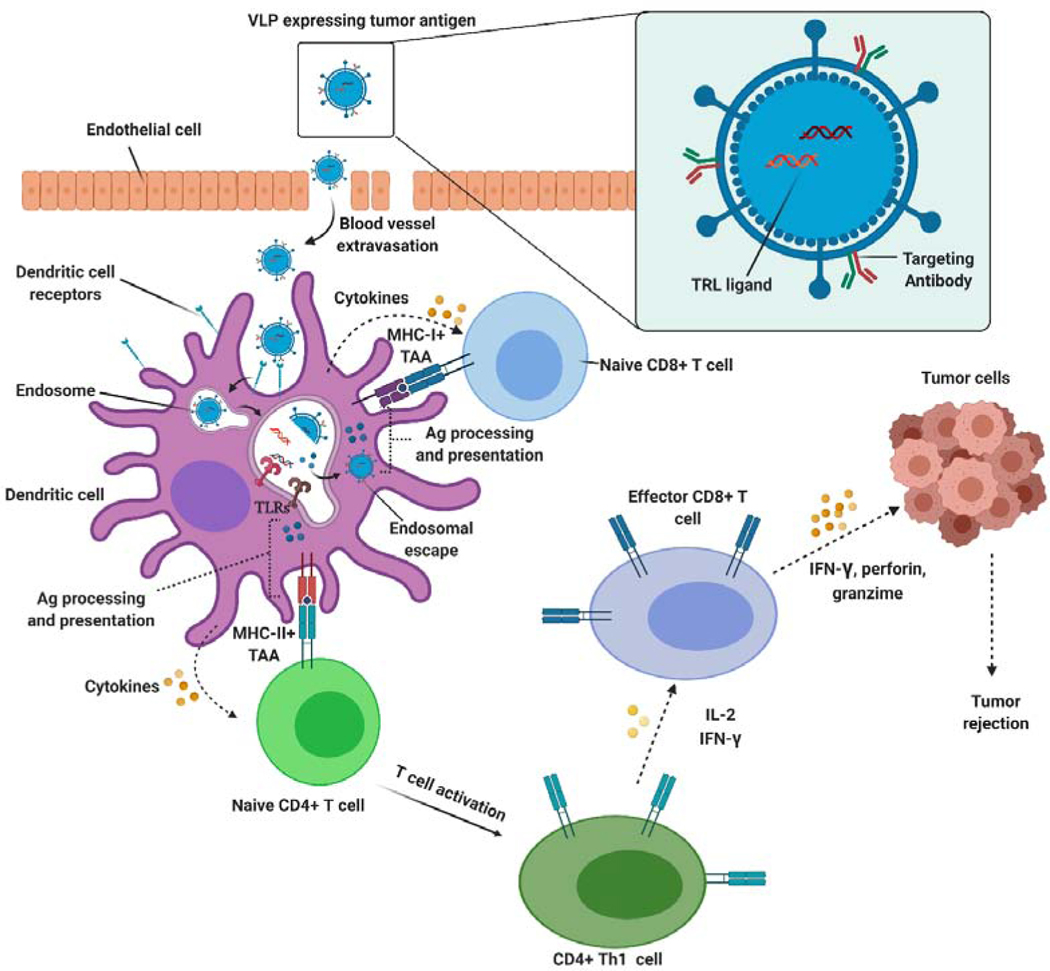
Figure 15.
Schematic of virus-like particle (VLP)-based vaccine’s cellular immune response against cancer. VLPs can cross blood vessels and be captured by antigen presenting cells such as dendritic cells (DCs) via micropinocytosis. Alternatively, VLPs can target DCs and be taken up via receptor-mediated endocytosis. Following uptake, VLPs are degraded and release the antigen and adjuvant. The adjuvant can be recognized by pattern recognition receptors such as toll-like receptors (TLRs). TLRs stimulate the maturation of DCs, upregulate co-stimulatory molecules (CD40, CD80/86) and secrete cytokines. The delivered cancer antigens are processed and presented as major histocompatibility complexes (MHCs) to T cells. The presented antigens enable activation of CD4+ T helper and CD8+ T lymphocytes. Upon activation and recognition of tumor cells, cytotoxic T lymphocytes induce cancer cell death via different pathways, including perforin/granzyme B, Fas-Fas ligand interaction, or the release of pro-inflammatory cytokines.

