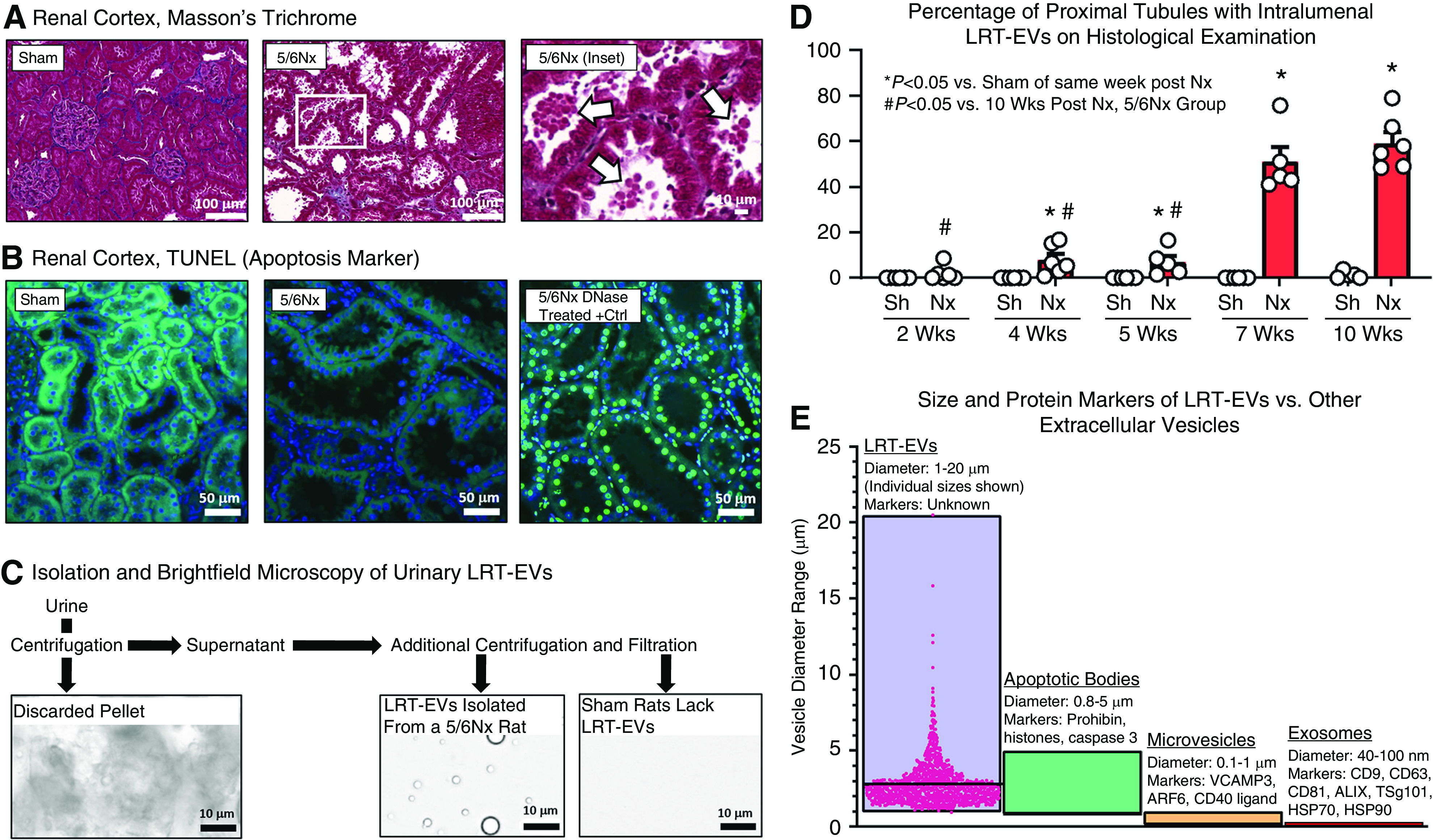
Figure 1.

Large renal tubular extracellular vesicles (LRT-EVs) spontaneously form in 5/6Nx but not sham-operated rats, are easily visible upon routine histological assessment, and are urine-excreted. (A) Examination of renal cortical tissue collected from 5/6 nephrectomy (5/6Nx) and sham-operated controls 10 weeks postsurgery. 5/6Nx rats exhibited extensive brush-border loss and the presence of large renal tubule extracellular vesicles (LRT-EVs) in proximal tubule lumens (arrows). (B) Terminal deoxynucleotidyl transferase–mediated digoxigenin-deoxyuridine nick-end labeling (TUNEL) indicates apoptosis by labeling DNA double-strand breaks. Kidney sections from sham-operated (sham; left) and 5/6Nx (center) rats were TUNEL negative (488 nm autofluorescence, green; 4′,6-diamidino-2-phenylindole [DAPI], blue). Importantly, the large vesicles within the lumen and the nuclei of the cells producing the vesicles were both TUNEL negative. A section treated with DNase (right) serves as a positive control for the assay (DAPI in blue colocalized with TUNEL in bright green). (C) These vesicles were isolated from 5/6Nx rat urine through a sequence of filtration and centrifugation (see Supplemental Figure 1 for detailed methodology). Sham rat urine lacked LRT-EVs. (D) Proximal tubules were scored for the presence of LRT-EVs in separate cohorts of sham and 5/6Nx rats at 2, 4, 5, 7, and 10 weeks postsurgery. Each dot represents one animal. (E) The size, as measured by microscopic analysis, and lack of apoptotic markers suggest these vesicles (LRT-EVs) represent a unique category of urine-excreted, extracellular vesicle, prompting proteomic analysis to characterize their composition. A subset of published diameters and protein markers of exosomes, microvesicles, and apoptotic bodies are represented here (5,14,15). Individual LRT-EV diameter measurements are shown; horizontal bar equals average. Nx, 5/6 Nx; Sh, sham.
