
Figure 2.
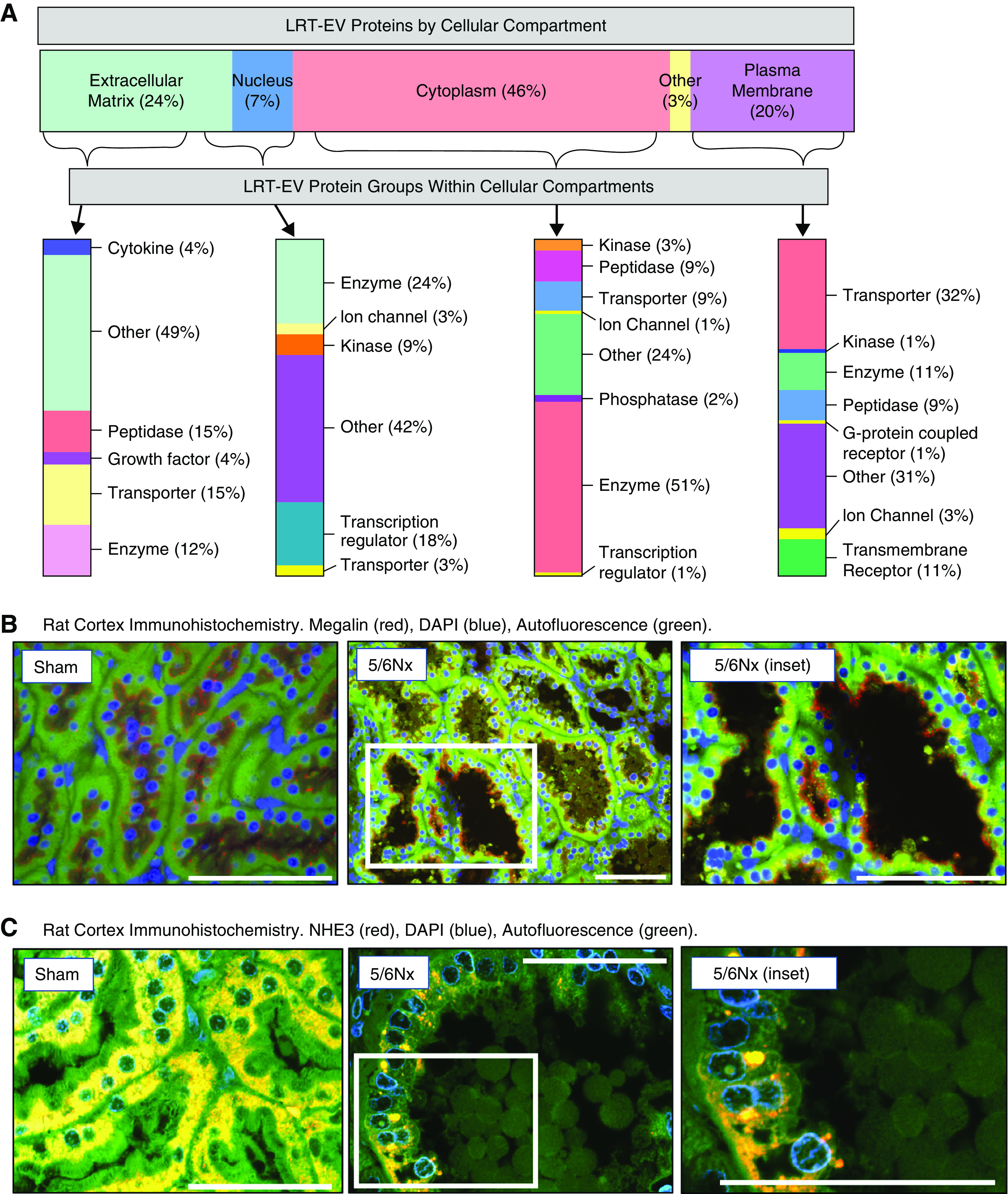
Proteomic analysis revealed LRT-EVs to contain proteins derived from numerous cellular compartments and of variable functional class. (A) Proteins identified through tandem mass spectrometry data analysis (N=3 with two technical replicates of each; six runs total), and meeting a minimum scan count of six and peptide count of two, were uploaded to Ingenuity Pathway Analysis to categorize these proteins based on cellular compartment and protein type/function. The percentage of identified LRT-EV proteins per cellular compartment (location) is depicted on the horizontal bar at the top. The proteins within each cellular compartment are then subcategorized by type, with percentage within each compartment indicated at the right of the vertical bars. (B) Proteomic analysis identified the plasma membrane transporter megalin in LRT-EVs. Representative immunohistochemistry of kidney tissues collected 10 weeks postsurgery show presence of megalin (red) localized at the base of the brush border on the apical membrane in sham-operated (sham) rats. In 5/6Nx rats, megalin can be seen in LRT-EVs that are within the tubule lumen and LRT-EVs emerging from the proximal tubule cells. The distribution of megalin is diffuse (yellow) or absent in some tubular cells. Image on right is an inset from center image. DAPI, blue; autofluorescence at 455 nm, green. Scale bar, 100 μm. (C) Proteomic analysis failed to detect sodium-hydrogen antiporter 3 (NHE3) in LRT-EVs. Immunohistochemistry of kidney tissue collected 10 weeks postsurgery shows the presence of NHE3 (red) in sham and 5/6Nx rat proximal tubule epithelial, but not in LRT-EVs. DAPI, blue; autofluorescence at 455 nm, green. Scale bar, 50 μm.