Fig. 4.
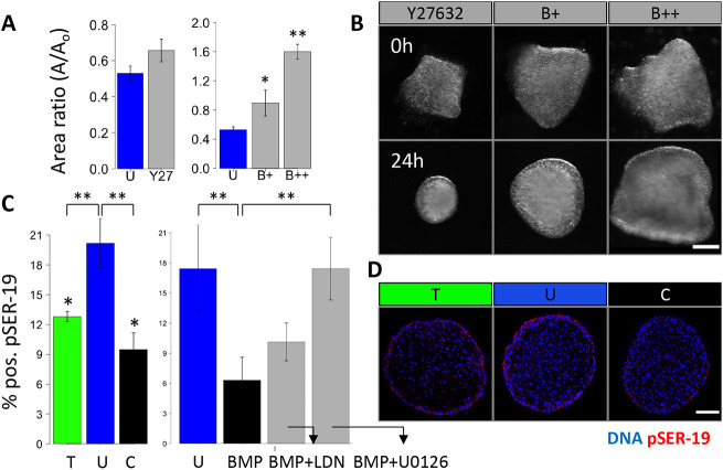
NM-myosin-II is inhibited in tissue growth driven by BMP and by compressive stress. (A) Compaction of HH25 AV cushions cultured with the ROCK inhibitor Y27632, 1 μM NM myosin II inhibitor blebbistatin (B+) and 10 μM blebbistatin (B++) for 24 h. (B) Representative images of cushions treated with blebbistatin showing varying degrees of compaction and rounding. (C) Percentage of pSER19-positive cells in HH25 cushions cultured under osmotic stress, and with BMP, the BMP inhibitor LDN and the MEK inhibitor U0126 for 24 h. (D) Representative immunofluorescence images stained for pSER19 of HH25 cushions cultured under osmotic stress over 24 h. Scale bars: 50 μm, n=4-6 cushions per condition per three independent experiments, data are mean±s.e.m., *P<0.05, **P<0.01 (ANOVA with Tukey's post-hoc test).

