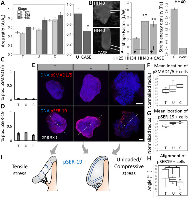
Fig. 7.
Compressive/tensile stress-regulated tissue compaction is conserved across stages of valve maturation and its interaction with extracellular matrix preserves leaflet morphology. (A) The compaction behavior of cushions across mid to late stages under osmotic stress conditions, and across late stage (HH40) compaction with collagenase II treatment (300 U). (B) Wide-field images of a HH40 leaflet before and after 24 h treatment with collagenase II (CASE). Shape factors, measured as aspect ratio, of HH25 and HH34 cushions cultured for 24 h, and HH40 leaflets cultured with collagenase II for 24 h. n=4-6 cushions per condition per three independent experiments, data are mean±s.e.m. Strain energy densities for control/unloaded and collagenase treated HH40 leaflets, three or four cushions per condition, data are mean±s.d. Percentage of pSmad1- and pSmad5- (C), and pSER19- (D) positive cells in HH40 leaflets cultured under osmotic stress for 24 h, n=3 leaflets per condition, data are mean±s.d. (E) Representative 3D reconstructed immunofluorescence images stained for pSmad1 and pSmad5, and pSER19 of HH40 leaflets cultured under osmotic stress over 24 h. The mean normalized radii for pSmad1- and pSmad5- (F), and pSER19- (G) positive cells in HH40 leaflets cultured under osmotic stress, n=3 leaflets per condition. (H) Angle between aligned pSER19-positive cells and long axis (perpendicular to elongation direction), n=3. Lines within boxes indicate the median. The length of each box represents the interquartile range. The whiskers indicate the highest and lowest observed values (unless outliers, which are indicated by points). (I) Schematic of alignment of pSER19-positive cells in HH40 leaflets under compressive/tensile stress and unloaded conditions. Scale bars: 100 μm. *P<0.05, **P<0.01.

